Highlighted Citation: Profiling of metastasis by whole animal staining
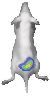
Recent advances in small animal imaging techniques allow in vivo whole-body detection of tumor cells tagged with fluorescence or luminescence. However, these techniques are still lacking in fine spacial resolution. Now, Kubota and colleagues report the application of new whole tissue clearing methods to allow immunofluorescence staining and imaging of whole organs and animals at the level of single cells.
The authors developed a combination of tissue clearing and imaging methods that they call “clear, unobstructed brain/body imaging cocktails and computational analysis” (CUBIC). Using CUBIC, they were able to image fluorescently-tagged metastatic tumor cells in a variety of tissues in mice, including challenging tissues like brain. CUBIC also allowed tumor cell imaging to be combined with 3-D immunofluorescence and nuclear staining using RedDot™2 Far-Red Nuclear Stain in whole mice and organs. Using CUBIC, the authors were able to bridge the resolution gap between in vivo imaging and traditional histological methods by imaging individual metastatic cells in whole organs and animals.
Kubota et al. (2017). Whole-Body Profiling of Cancer Metastasis with Single-Cell Resolution. Cell Reports 20, 236-250. http://dx.doi.org/10.1016/j.celrep.2017.06.010